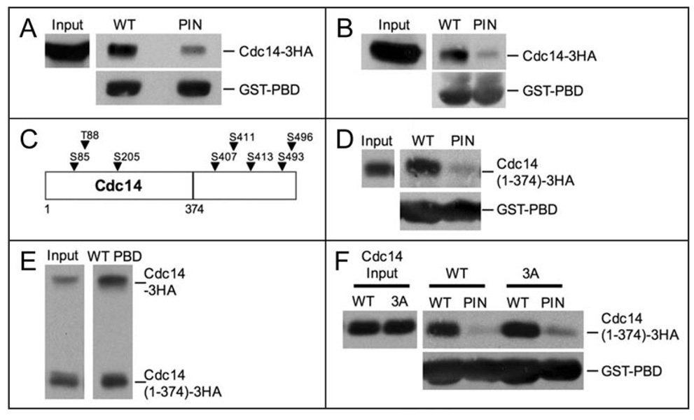
Figure 4.
The Polo-box domain (PBD) of Cdc5 binds to Cdc14. (A) Whole cell extracts from cells carrying Cdc14-3HA fusion (A1411) were incubated with WT or PIN PBD-GST fusions immobilized on glutathione (GSH) beads. Western blots show the amount of Cdc14-3HA in whole cell extracts (Input, top left), the amount of Cdc14-3HA coprecipitated with WT or PIN PBD-loaded GSH beads (top right), and the amount of WT or PIN PBD-GST fusion immobilized on GSH beads (bottom). (B) Lysates from cells carrying a Cdc14-3HA fusion (A1411) were prepared under denaturing conditions as described in Materials and methods. WT or PIN PBD-GST fusions immobilized on GSH beads were incubated with “denatured” extract. Western blots show the amount of Cdc14-3HA in “denatured” extracts (Input, top left), the amount of Cdc14-3HA coprecipitated with WT or PIN PBD-loaded GSH beads (top right), and the amount of WT or PIN PBD-GST fusion immobilized on the GSH beads (bottom). (C) Schematic diagram of Cdc14 showing the locations of putative PBD binding sites (arrows). The line denotes where the Cdc14(1–374) truncation ends. (D) WT or PIN PBD-GST fusions immobilized on GSH beads were incubated with “denatured” extract from cells carrying a Cdc14(1–374)-3HA fusion (A19448). Western blots show the amount of Cdc14(1–374)-3HA in “denatured” extracts (Input, top left), the amount of Cdc14(1–374)-3HA pulled-down with WT or PIN PBD-loaded GSH beads (top right), and the amount of WT or PIN PBD-GST fusion immobilized on the GSH beads (bottom). (E) WT PBD-GST fusions immobilized on GSH beads were incubated with “denatured” extract from diploid cells carrying a Cdc14-3HA and a Cdc14(1–374)-3HA fusion (A19422). Western blots show the amount of Cdc14-3HA and Cdc14(1–374)-3HA in “denatured” extracts (Input, left), the amount of Cdc14-3HA and Cdc14(1–374)-3HA coprecipitated with WT PBD-loaded GSH beads (right). (F) WT or PIN PBD-GST fusions immobilized on GSH beads were incubated with “denatured” extract from cdc14Δ cells carrying Cdc14(1–374)-3HA (A21384) or Cdc14(1–374, 3A)-3HA fusions (A21386) on centromeric plasmids. Western blots show the amount of Cdc14(1–374)-3HA and Cdc14(1–374, 3A)-3HA in “denatured” extracts (Input, top left), the amount of Cdc14(1–374)-3HA and Cdc14(1–374, 3A)-3HA precipitated with WT or PIN PBD-loaded GSH beads (top right), and the amount of WT or PIN PBD-GST fusion immobilized on the GSH beads.

