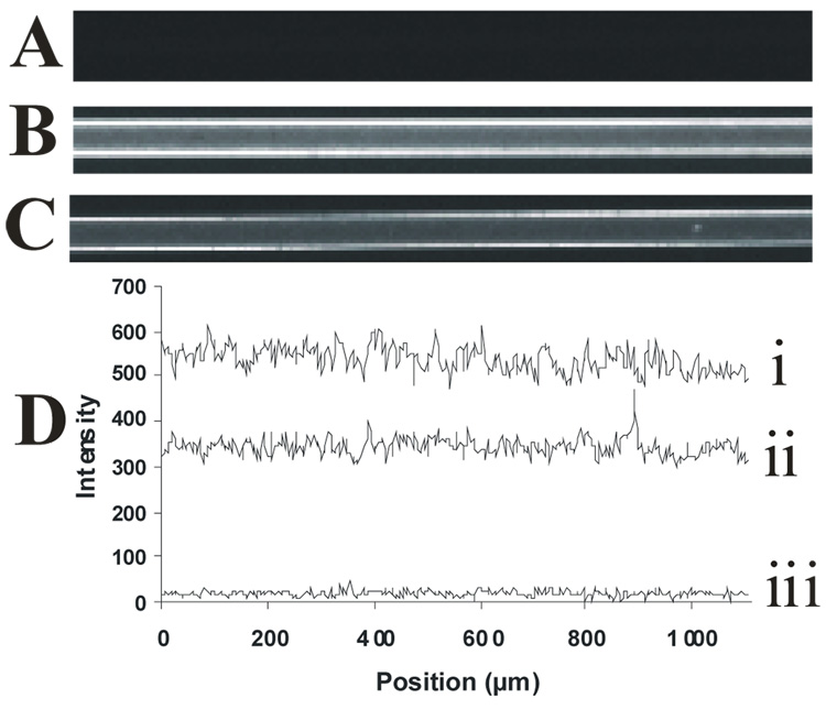
Figure 4.
Uniformity of PLB coatings formed in situ. Fluorescence images were collected following introduction of the fluorogenic membrane stain, FM 1–43, into (A) bare capillary, (B) DOPC-coated capillary, and (C) poly(bis-SorbPC)-coated capillary. (D) Line scans obtained from regions along the capillary wall, showing the relative fluorescence intensities as a function of position along the capillary i) DOPC capillary, ii) bis-SorbPC capillary, iii) bare capillary. All capillaries are 50 µm i.d.

