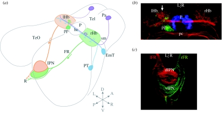
Figure 2.
Asymmetric habenular circuitry in zebrafish. (a) Schematic showing connectivity of the habenular complex in larval zebrafish. A significant afferent input derives from migrated neurons of the eminentia thalami, which is thought to form the entopeduncular/peripeduncular complex in adult zebrafish (note that the teleostean entopeduncular complex is not part of the pallidum and does not correspond to the EP of amniotes; Wullimann & Mueller 2004). EmT neurons project bilaterally, innervating both left and right habenulae. A subset of left- and right-sided neurons in the anterior pallium are a source of asymmetric innervation, selectively terminating in a small medial domain of the right habenula (indicated in purple). In addition, a small afferent input may derive from the posterior tuberculum (Hendricks & Jesuthasan 2007). In the epithalamus, the left-sided parapineal exclusively innervates the left habenula (Concha et al. 2003). Habenular neurons project efferent axons that course in the fasciculus retroflexus. A major target is the interpeduncular nucleus: left- and right-sided axons are segregated along the dorso-ventral (DV) axis of the IPN in a laterotopic manner (Aizawa et al. 2005). A smaller and apparently symmetric contingent of habenular axons terminates caudal to the IPN in the serotonergic raphe. (b) Neuroanatomical asymmetries in the dorsal diencephalon. Anti-acetylated tubulin immunostaining (red) shows that the left habenula contains a greater density of neuropil, especially in the dorsomedial aspect of the nucleus. The pineal (blue) and parapineal (green) are visualized by the expression of green fluorescent protein (GFP) in a Tg(foxD3:GFP) transgenic larva. The parapineal is asymmetric in both its location and connectivity, and its efferent axons preferentially terminate in the asymmetric medial neuropil of the left habenula. Dorsal view, anterior top. (c) Three-dimensional confocal reconstruction showing habenular axon terminals in the ventral midbrain labelled using lipophilic tracer dyes applied to the habenulae. Left-sided axons were labelled with DiD (red) and right-sided axons with DiI (green). The dorsal IPN is almost exclusively innervated by left-sided axons, whereas the ventral target receives a majority of right-sided inputs. Dorsal view, anterior top. Tel, telencephalon; EmT, eminentia thalami; PT, posterior tuberculum; Pa, pallium; sm, stria medullaris; Hb, habenula; hc, habenular commissure; pp, parapineal, P, pineal; pc, posterior commissure; FR, fasciculus retroflexus; TeO, optic tectum; IPN, interpeduncular nucleus; a, anterior; p, posterior; l, left; r, right; d, dorsal; v, ventral. Adapted from Bianco et al. (2008). A number of these asymmetry phenotypes are conserved in the distantly related teleost medaka (Oryzias latipes; Signore et al. 2009).

