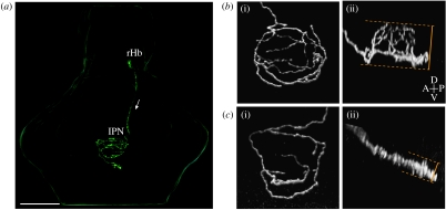
Figure 3.
Zebrafish Hb–IPN projection neurons elaborate one of two distinct axon terminal arbour morphologies. (a) Three-dimensional reconstruction showing a single right habenular projection neuron that was labelled by focal electroporation with a construct driving the expression of membrane GFP, in an intact larval zebrafish brain (4 days post-fertilization). The cell body, located in the right habenula (rHb) extends an axon down the right FR (indicated by arrow) that terminates in the IPN. Habenular neurons elaborate remarkable axon arbours within the IPN that cross the ventral midline multiple times. Scale bar, 100 μm. (b(i),c(i)) Dorsal and (b(ii),c(ii)) lateral confocal reconstructions of single habenular axon arbours in the IPN. (b(i),(ii)) Example of an L-typical axon arbour, formed by 84% of left habenular neurons. These arbours are located in the dorsal IPN and are shaped similar to a domed crown and arborize over a considerable dorsoventral extent (compare dorsal (b(i)) and lateral (b(ii)) views of an example L-typical arbour). (c(i),(ii)) Example of an R-typical axon arbour, which is considerably flatter, localized to the ventral IPN and formed by 90% of right habenular neurons. Adapted from Bianco et al. (2008).

