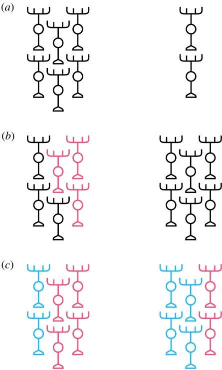
Figure 4.
Models for lateralization of neural tissue. (a) Equivalent regions on the left and right of the CNS are identical in composition and differ only in overall size. (b) Unique types of neuron, or patterns of connectivity, may be specified on either the left or right or both sides (indicated by unique red neurons on the left in this schematic). (c) Identical circuit components might exist on both sides of the CNS, but in different ratios. Note that these models are in no way mutually exclusive. In fact, it is likely that all three strategies may be involved in the lateralization of DDC circuitry (see the main text).

