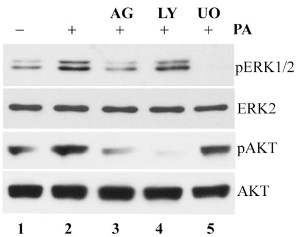
FIGURE 2.
EGFR-dependent ERK1/2 and PI3K activation in P. aeruginosa–infected HUCL cells. HUCL cells were pretreated with 500 nM AG1478 (lane 3), 10 μM LY294002 (lane 4), or 10 μM U0126 (lane 5) for 30 minutes and stimulated with P. aeruginosa at a cell-to-bacterium ratio of 1:25 in the presence of the inhibitors for 1 hour. Uninfected cells (lane 1) and cells infected with P. aeruginosa alone (lane 2) were used as controls. Cell lysates (10 and 30 μg) were used for detection of phospho-ERK1/2 (pERK1/2) and phospho-Akt (pAKT), respectively. To normalize protein loading and determine the change of ERK2 and Akt in infected cells, anti-ERK2 (ERK2) and anti-Akt (AKT) were used to probe the samples.

