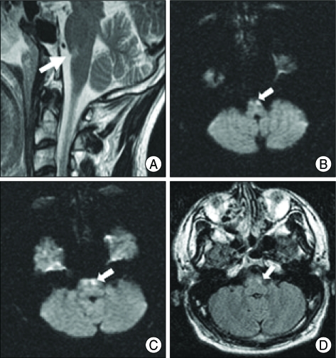
Fig. 2.
A : Sagittal T2-weighted magnetic resonance (MR) image of the cervical spine showing focal high signal intensity in the left lower pons (arrow). B : Axial diffusion-weighted MR image obtained at the 2nd hospital day after injury. MR images demonstrated a focal hyperintensity of the ventral upper medulla (arrow), more prominent in the left side. C : Axial diffusion-weighted MR displaying a focal hyperintensity in the lower pontine lesion (arrow). D : Axial fluid-attenuated inversion recovery MR image showing a focal hyperintensity in the ventral pontomedullary junction.

