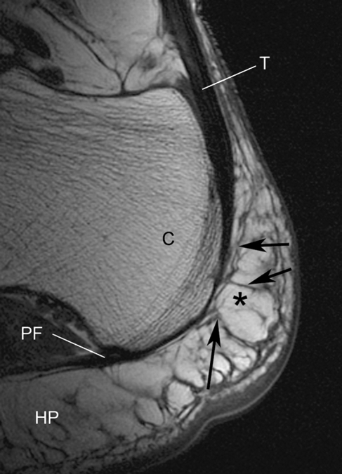
Fig. 2.
A non-fat suppressed sagittal MR image of the normal heel of a young female volunteer (age 42 years) showing how the Achilles tendon (T) is directly continuous with the plantar fascia (PF) around the posterior aspect of the calcaneus (C). Fat (asterisk) is high signal (bright). Tendons, fascia and septa are low signal (black). Note how the heel fat pad (HP) extends upwards behind the tendon and that its fibrous septa are connected to it (arrows).

