Fig. 5.
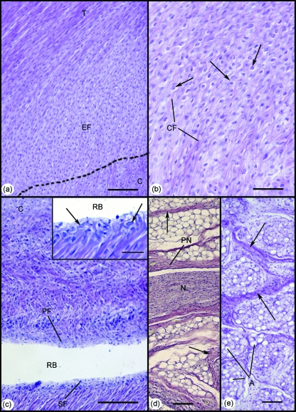
Development of the enthesis organ in the 332-mm foetus. (a) The enthesis fibrocartilage (EF) was first identifiable at this stage. Note the gradual bending of collagen fibres, as the Achilles tendon (T) inserts into the cartilaginous calcaneus (C). Scale bar: 100 µm. (b) Higher power view of the enthesis fibrocartilage in (a). Note the characteristic rounded fibrocartilage cells (arrows) interspersed between small bundles of collagen fibres (CF). Scale bar: 50 µm. (c) Both the sesamoid (SF) and periosteal (PF) fibrocartilages were also evident in the 332-mm foetus – forming the walls of the distal part of the retrocalcaneal bursa (RB). C, calcaneus. Scale bar: 100 µm. Inset: Higher power view of the sesamoid fibrocartilage in (c). Note the characteristic rounded cells (arrows). Scale bar: 100 µm. (d) Large myelinated nerve fascicles (N) surrounded by a prominent perineurium (PN) were evident within Kager's fat pad at this stage. Collections of well-differentiated adipocytes were separated by thick fibrous septa (arrows). Scale bar: 100 µm. (e) At this stage of development, adipogenesis was evident for the first time in the heel pad. Note the organisation of adipocytes (A) into locules, separated by thick fibrous septa (arrows). Scale bar: 100 µm. All slides were stained with Hansen's hematoxylin.

