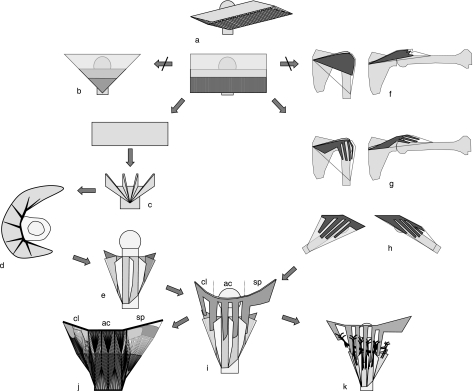
Fig. 3.
Generic model of deltoid. A unipennate muscle (a) is stepwise transformed into a deltoid model (i). Left branch: generic model of ET. Right branch: OT. For further explanations, see text. The final model (i) shows bipennate OT lamellae interdigitating with bipennate ET blades. cl, ac, sp: clavicular, acromial and spinal parts, resp. j: 2D rendition of muscle fiber structure in a deltoid with seven ET blades and one clavicular, three acromial and one spinal OT lamellae. The tendon structure is detailed in Fig. 6. k: The inter-lamella spaces are natural pathways for segmental neurovascularization.

