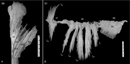
Fig. 5.
Overview of deltoid origin and end tendons. (A) (specimen S6) ET blades, superimposed, anterior view. This specimen had seven ET blades. The anterior five ET blades are visible (b1–b5); the posterior two blades are hidden beneath the others (see Fig. 6). (B) (specimen S1) Origin tendons, front view, amputated in one piece at origin line after removal of muscle fibers. cl, ac, sp: clavicular, acromial and spinal parts, respectively, marked by white dashed lines. This specimen had two superimposed clavicular OT lamellae (superficial lcs, deep lcd); four acromial lamellae (la1–la4); and one spinal lamella (ls). The posterior spinal OT sheet (ss) is not lamellated. It is superficial to the muscle, with muscle fibers arising unipennately from the backside, as shown by the posterior deltoid muscle mass left in situ. The black dashed line marks the posterior border of the bipennate part of the spinal lamella, which is posterior continuous with the superficial spinal OT sheet.

