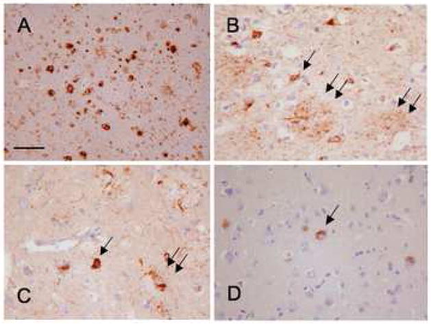
Figure 1.
A: Aβ immunohistochemistry showing numerous mature and diffuse plaques in the superior temporal cortex.
B: The AT8 anti-tau antibody shows numerous neurofibrillary tangles (arrow), neuropil threads and plaque-associated abnormal neurites (double arrow) in the same area.
C: Immunohistochemistry with the anti-4R-tau antibody, RD4, shows a similar staining pattern.
D: The anti-3R-tau antibody (RD3) mostly stained neurofibrillary tangles. Bar on A represents 100 mm on A and 50 mm on B–D.

