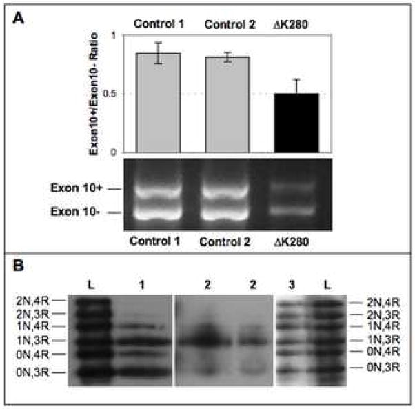
Figure 2.
A: RT-PCR analysis of mRNA from the frontal cortex of the MAPT ΔK280 case and two healthy controls. In the top panel, the bars depict the ratios of the exon 10+ (4R-tau) and exon 10− (3R-tau) transcripts as quantified by intensities of the respective bands (see lower pane). Error bars represent standard error of the mean.
B: Western blot analysis of dephosphorylated, soluble (Lane 1) and sarkosyl-insoluble (Lanes 2) tau from the frozen frontal cortex of the ΔK280 case. Lane 3 shows sarkosyl-insoluble tau from the frontal cortex of another Alzheimer’s disease case, without MAPT mutations. Lanes L are recombinant tau isoforms, as labelled on the left and right edges.

