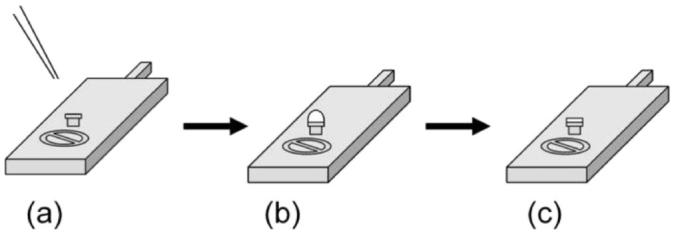
Figure 1.

(a) Initially, a drop of the crystalline mesophase is deposited on a clean, empty rivet that is held in place on the cryo-sample stage at room temperature. (b) The sample stage with the crystalline mesophase is immediately frozen in slushed liquid nitrogen and transferred under vacuum to the cryo-SEM. (c) The sample is subsequently fractured to reveal a clean surface that is then sublimed and sputter coated with platinum prior to imaging.
