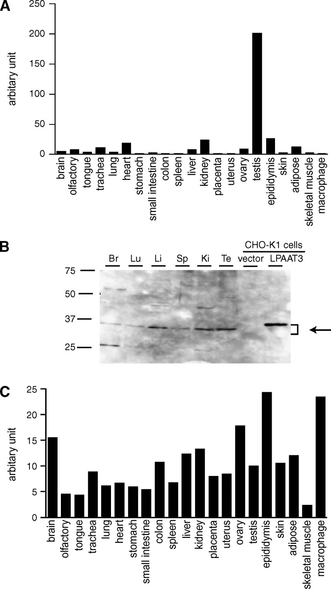
Fig. 2.
Expression profile of mLPAAT3 and mMBOA7 (LPIAT1) in mice. Expression levels of mLPAAT3 mRNA (A) and mMBOA7 (LPIAT1) mRNA (C) in 21 tissues from C57BL/6J mice were analyzed using quantitative real-time PCR. mLPAAT3 (A) was expressed predominantly in the testis, whereas mMBOA7 (LPIAT1) (C) was ubiquitously expressed. Similar results were obtained in a separate independent experiment. B: Expression of mLPAAT3 was analyzed at the protein level by Western blots using anti-mLPAAT3 antiserum. Three micrograms of 100,000 g pellets from various tissues were loaded in each lane. Br, Lu, Li, Sp, Ki, and Te stand for brain, lung, liver, spleen, kidney, and testis, respectively. mLPAAT3 was highly expressed in the testis. High expression was noted in the liver and kidney as well. The results are representative of three independent experiments.

