Fig. 6.
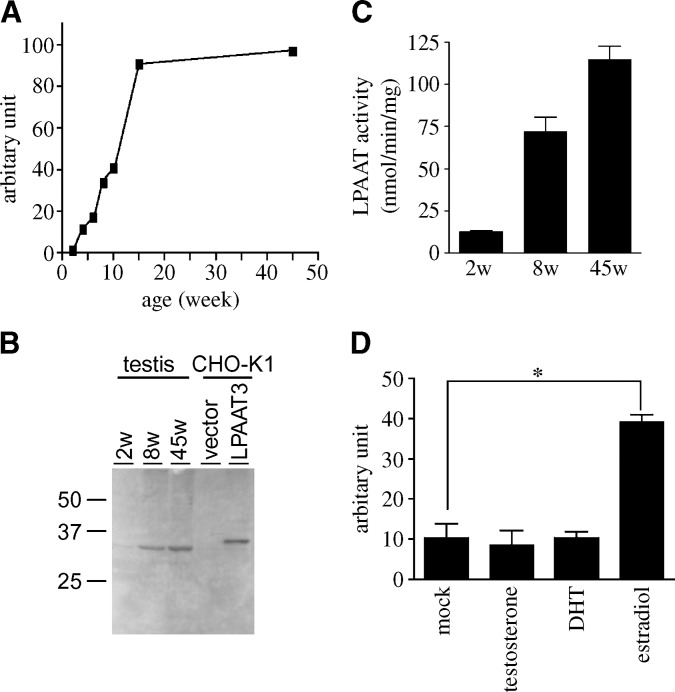
Age-dependent expression of mLPAAT3 in the testis and mLPAAT3 induction in testicular cell line. A: mLPAAT3 mRNA expression in the testis at various ages was compared using real-time quantitative PCR. mLPAAT3 mRNA expression is enhanced significantly in an age-dependent manner until 15 weeks of age. The results are representative of two independent experiments. B: mLPAAT3 protein expression in the testis at different ages was compared by Western blots using anti-mLPAAT3 antiserum. Four micrograms each of 100,000 g pellets at different ages were loaded. Results are representative of two independent experiments. C: LPAAT activity of 2, 8, and 45 weeks testis was examined using 1 μg protein (100,000 g pellet), 25 μM arachidonoyl-CoA (33,000 dpm), and 50 μM palmitoyl LPA. Data represent mean + SD of triplicate samples measurements. The results are representative of two independent experiments. D: Testicular cell line TM4 cells were treated with either mock, 100 nM β-estradiol, dihydrotestosterone (DHT), or testosterone for 24 h. mLPAAT3 mRNA level was compared using real-time quantitative PCR. β-Estradiol induced mLPAAT3 significantly. Data represent mean + SD of three independent experiments. Statistical significance was analyzed using ANOVA with Tukey post hoc pairwise comparisons. *P < 0.05.

