Fig. 7.
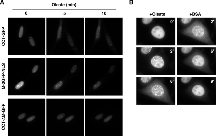
Oleate promotes domain M-dependent export of CCT-GFP. A: CHO58 cells expressing CCT-GFP, CCT-ΔM-GFP, or M-2GFP-NLS were prepared for live-cell imaging as described in Materials and Methods. Cells were mounted on a 37°C heated stage and treated with an oleate-BSA complex (300 μM oleate), and images were captured (750 ms exposures) at the indicated times. B: CHO58 cells expressing CCT-GFP were treated with oleate (320 μM complexed with BSA) at 40°C, and images were captured (150 ms exposures) at 30 s intervals for 6 min. Cells that displayed significant nuclear export of CCT-GFP were identified, and medium was replaced with prewarmed (40°C) oleate-free F-12 medium containing 0.2% BSA. Images were captured for a further 9 min. The Quicktime movie from which these images were taken is shown in supplementary Fig. III.

