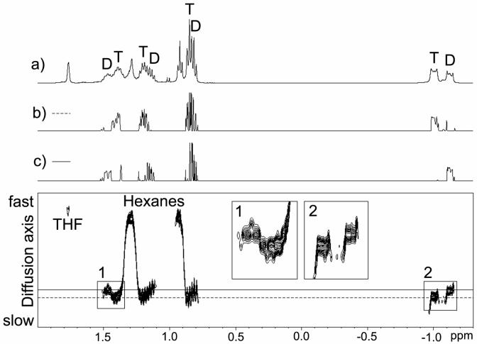
FIGURE 1.
1H DOSY spectrum of n-BuLi in THF-d8 at -84 °C. (a) 1H spectrum at 187 K, the labeled butyl resonances of dimeric (D) and tetrameric (T) n-BuLi were assigned by 2D-TOCSY. (b) 1D Slice of the DOSY spectrum at the diffusion coefficient of the tetramer (- - -). (c) 1D Slice of the DOSY spectrum at the diffusion coefficient of the dimer (-).

