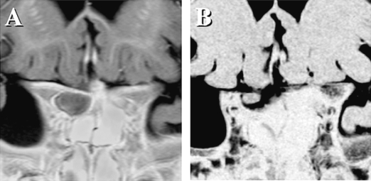
Fig. 2.
A coronal image shows a swollen optic nerve in the right optic canal (A; multiple planner reconstruction image). Due to deterioration of visual acuity, the optic canal was unroofed to decompress the affected nerve. After cycles of chemotherapy, the optic nerve gradually shrank (B; three-dimensional MR cisternography).

