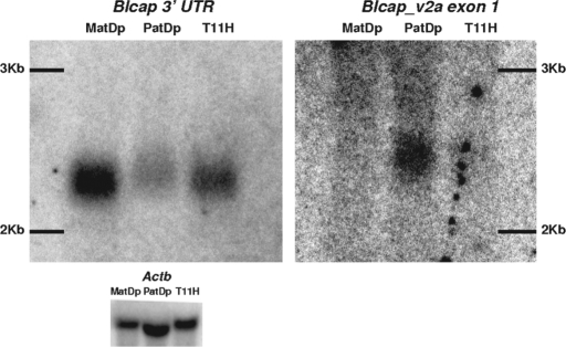
Figure 4.
Detection of Blcap_v1a and Blcap_v2a in MatDp(dist2), PatDp(dist2) and non-UpDp (T11H) newborn brain RNA by northern hybridization. The signal obtained with an Actb probe is shown as a loading control (bottom). Left: Hybridization signal produced by the probe designed to the last Blcap exon (N1 in Fig. 1B). A single band was observed in all three lanes where the relative band intensity was highest in the MatDp(dist2) lane and lowest in the PatDp(dist2) lane. Band position and relative intensities were consistent with the known size of the major Blcap transcript Blcap_v1a (2079 bp) and overall preferential expression of Blcap from the maternal allele in newborn brain. Right: Results obtained with a probe specific to the first exon of Blcap_v2a (N2 in Fig. 1B). No band was present in the MatDp(dist2) lane. A single band was visible in the PatDp(dist2) lane. At the corresponding position in the non-UpDp lane, a very faint band was present. This indicates paternal-only expression of Blcap_v2a. The band was slightly shifted upward relative to the band obtained with the N1 probe, as is expected of the slightly larger Blcap_v2a (2258 bp). The exposure times necessary to visualize the results for Blcap_v2a were considerably longer than for the N1 probe, which is consistent with the low abundance of Blcap_v2a relative to Blcap_v1a as determined by qRT–PCR.

