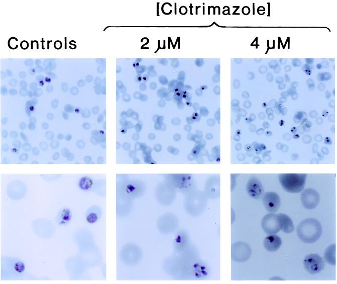
Figure 3.
Morphological appearance of P. falciparum cultures (A4 clone) after 24-hr incubation with CLT. Parasites were synchronized at the ring stage. Shown is the morphology of Giemsa-stained thin blood smears from drug-free control cultures (Left) and cultures incubated with either 2 μM CLT (Middle) or 4 μM CLT (Right) for 24 hr. The views are shown at two different magnifications: ×400 (Upper) and ×1,000 (Lower). Note parasite pyknotic changes and prevalence of ring forms in cultures treated with CLT.

