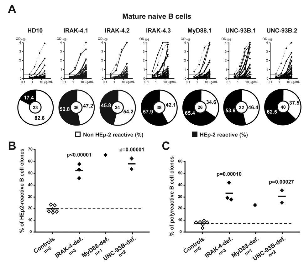
Figure 5. Defective peripheral B cell tolerance checkpoint in IRAK-4-, MyD88- and UNC-93B-deficient patients.
(A) Increased frequency of HEp-2-reactive antibodies in IRAK-4-, MyD88-, and UNC-93B-deficient mature naïve B cells. Antibodies from mature naïve B cells from one control (HD10), three IRAK-4-deficient patients, one MyD88-deficient, and two UNC-93B-deficient patients were tested in ELISA for reactivity with HEp-2 cell lysate. Dotted lines show ED38-positive control (Meffre et al., 2004; Wardemann et al., 2003). Horizontal lines show cut-off OD405 for positive reactivity. For each individual, the frequency of HEp-2-reactive (in black) and non HEp-2-reactive (in white) clones is summarized in pie charts with the number of antibodies tested indicated in the centers. The frequency of (B) HEp-2-reactive and (C) polyreactive (tested against single-stranded DNA, double-stranded DNA, insulin and lipopolysaccharide) clones in mature naive B cells of IRAK-4-, MyD88- and UNC-93B-deficient patients is higher than in controls. Each diamond represents an individual, and the average is shown with a bar. Statistically significant differences between patients and controls are indicated.

