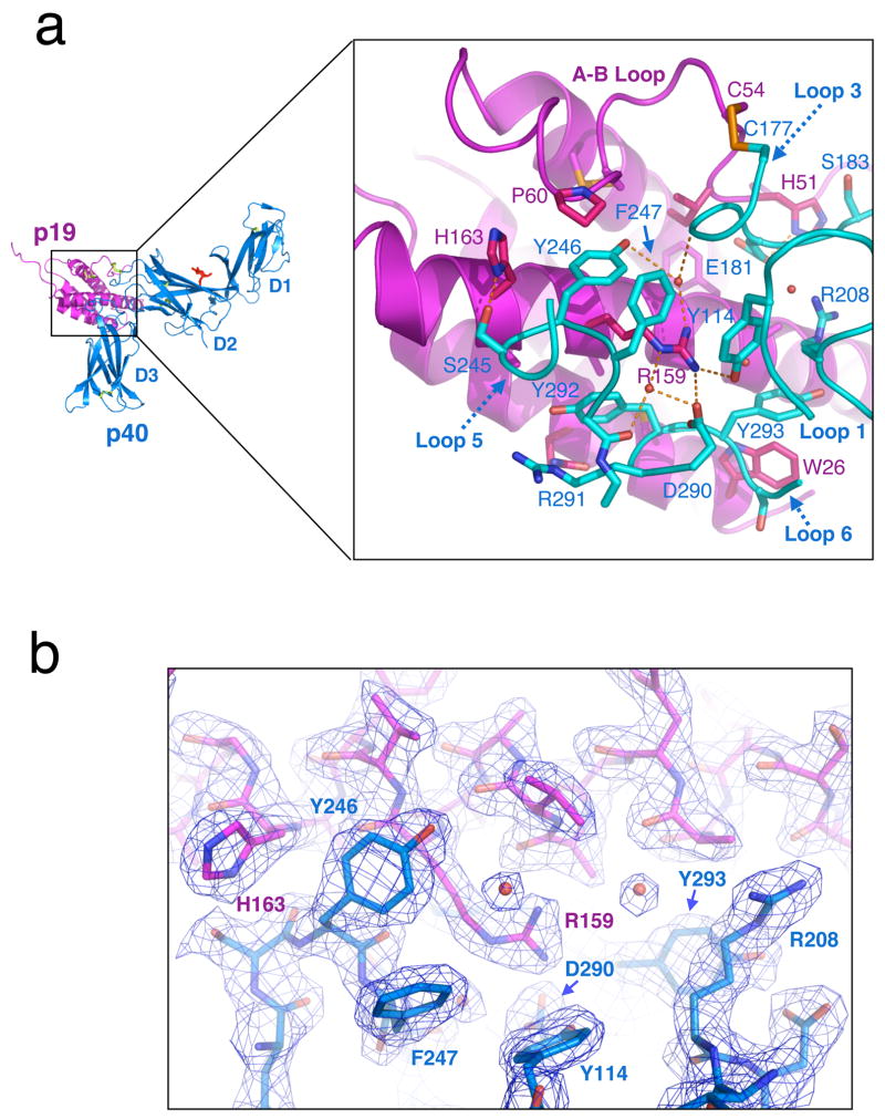
Fig. 3.
Structural anatomy of the p19–p40 interface. (a) Close-up view of the secondary structure and amino acid contacts between p19 and p40. (b) Cut away view of the ‘Arginine pocket’ 2Fo-Fc electron density contoured at 1.5 σ. Several well-ordered waters are visible at the interface, stabilizing the interaction of p40 pocket residues with Arg159.

