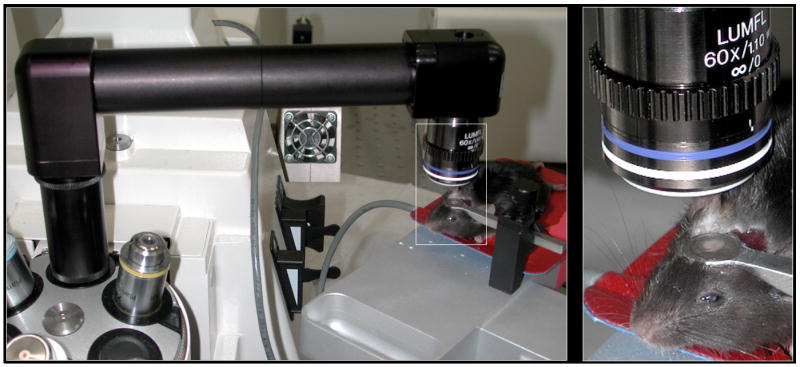
Figure 1. Optical Imaging in vivo of the mouse parietal cortex.

Left panel shows the objective inverter attached to a Zeiss LSM 510 multiphoton microscope positioning the 60x 1.1 NA water immersion objective above the mouse parietal cortex. Right panel is a higher magnification of the stainless steel ring holder that is glued to the skull and immobilizes the brain. The center ring has been filled with 2 % agarose (Sigma type VII) and sealed from above with a glass coverslip (#0). This essentially eliminates motion artifacts due to breathing when the hole in the cranium is less than 1-2 mm in diameter. The red heating pad is maintained at 37°C.
