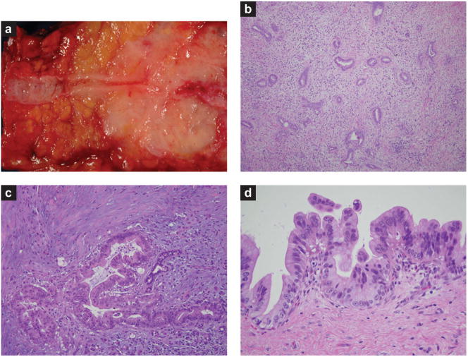
Figure 1.
Pathology of pancreatic adenocarcinoma and its precursor lesions. (a) Gross photograph of an infiltrating adenocarcinoma. Note the dramatic narrowing of the pancreatic duct associated with the poorly defined white neoplasm. (b) Low-power photomicrograph of an infiltrating adenocarcinoma. Note the haphazard arrangement of the glands and the associated non-neoplastic desmoplastic stroma. (c) High-power photomicrograph of an infiltrating adenocarcinoma. Note the desmoplastic stroma and the marked pleomorphism in the cancer relative to the trapped non-neoplastic duct. (d) High-grade pancreatic intraepithelial neoplasia.

