Figure 5.
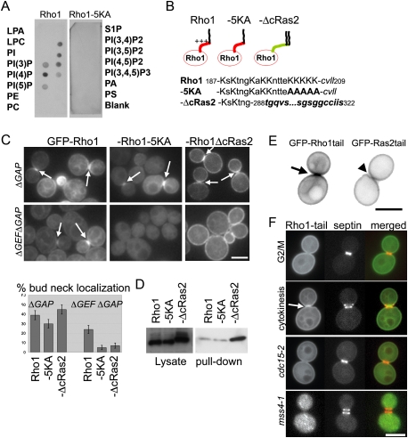
The Rho1 PBS binds lipids and is a bud neck targeting signal. (A) PIP strip (Echelon) membranes were incubated with yeast cell lysates from the cells expressing endogenous-level GFP-RHO1 or GFP-RHO1-5KA. Membrane-bound proteins were detected with an anti-GFP antibody. (LPA) Lysophosphatidic acid; (LPC) lysophosphatidylcoline; (PI) phosphatidylinositol; (PE) phosphatidylethanolamine; (PC) phosphatidylcholine; (S1P) sphingosine-1-phosphate; (PA) phosphatidic acid; (PS) phosphatidylserine. (B) Amino acid sequence of the Rho1 tail and derivatives. Positively charged residues (K) are in capital letters. The CAAX sequence of Rho1 and plasma membrane targeting sequence of Ras2 (288–322) are in italics. Mutated residues are in bold face. (C) Localization of Rho1 tail mutants. GFP-tagged Rho1 mutants were localized in the presence (ΔGAP) or absence (ΔGEFΔGAP) of Rho1 GEFs. Log-phase cells at 24°C were imaged without fixation. Bar, 3 μm. (D) The level of GTP-Rho1 was measured in the indicated strains by a RBD pull-down assay as in Figure 2B. (E) Localization of GFP-Rho1tail and GFP-Ras2tail during cytokinesis. (F) GFP-Rho1tail was expressed in the strain expressing septin (Shs1)-mRFP. The Rho1 tail localized to the bud neck only after the septin rings split. The localization of GFP-Rho1tail in cdc15-2-arrested cells and after inactivation of Mss4 (3 h, 37°C) are shown. Arrows indicate accumulation to the bud neck.

