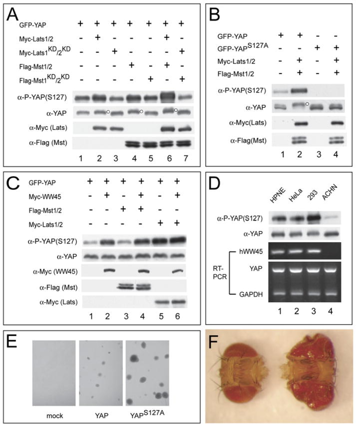
Figure 4. Delineation of a Mammalian Hippo Signaling Pathway Leading to YAP S127 Phosphorylation.
(A) HEK293 cells were transfected with epitope-tagged constructs as indicated. Note the increased YAP(S127) phosphorylation and mobility retardation resulting from Lats1/2 (lane 2), Mst1/2 (lane 4), or both (lane 6), but not the respective kinase-dead forms or their combinations (lanes 3, 5, and 7). The slower-migrating species of YAP was indicated by a small dot to the right of the protein band.
(B) HEK293 cells expressing the indicated plasmids were analyzed. The slower-migrating species of YAP was indicated by a small dot to the right of the protein band.
(C) ACHN cells expressing the indicated plasmids were analyzed. Note the failure of Mst1/2 (lane 3), but not Lats1/2 (lane 5), to induce YAP(S127) phosphorylation, and the rescue of this defect by hWW45 (lane 4). Also note that hWW45 alone (lane 2) can induce YAP(S127) phosphorylation.
(D) Various cell lines were probed with α-P-YAP(S127) and α-YAP, and analyzed by RT-PCR for the expression of indicated genes. Note the absence of hWW45 expression and the much diminished levels of YAP(S127) phosphorylation in ACHN cells (lane 4).
(E) HPNE cells stably expressing YAP (middle), but not those expressing an empty vector (left), grew in soft agar to form colonies. HPNE cells stably expressing YAPS127A grew to significantly larger colonies (right). Similar results were obtained with multiple independently established cell lines.
(F) Adult heads from flies expressing YAP (left) or YAPS127A (right) in the eye. The genotypes are: GMRGal4/UASYAP (left) and GMRGal4/UASYAPS127A (right). The heads were photographed together to show their relative size.

