FIGURE 1.
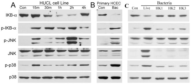
P. aeruginosa stimulates IκB-α degradation/phosphorylation in HUCL or primary HCE cells. HUCL (A) or primary HCE cells (B) were grown to ~90% confluence on 6-well plates and were serum-starved overnight. Cells were then infected with live P. aeruginosa at a cell-to-bacterium ratio of 1:50 (A and B) over a 4-hr time course. HUCL cells were also challenged with heat-killed P. aeruginosa (C) for 2 hr at different cell-to-bacterium ratio 1:50 (HK1), 1:100 (HK2), or 1:200 (HK3); Live, positive control with live P. aeruginosa (ratio 1:50). Cell lysates were prepared at the designated time points p.i. and 20 μg of protein was subjected to SDS-PAGE and immunoblotting using phospho-IκB-α (p-IκB-α), IκB-α, phospho-p38 (p-p38), and phospho-JNK (p-JNK), with anti-p38 and JNK as loading control. These results are representative for three independent experiments.

