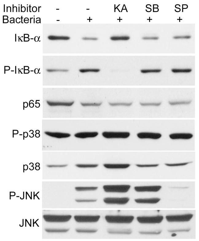
FIGURE 4.
Effects of inhibitors on P. aeruginosa–induced signal activation. HUCL cells were preincubated with 25 μM kamebakaurin, 10 μM SB203580, or 0.5 μM SP600125 for 30 min and infected with P. aeruginosa at a cell-to-bacterium ratio of 1:50 for 2 hr. Cells were then lysed and subjected to Western blotting analysis with anti-phospho-IκB-α (p-IκB-α), IκB-α, p65 of NF-κB (p65), phospho-p38 (P-p38), and phospho-JNK (p-JNK), with anti-p38 and JNK as loading controls. Results are representative of two independent experiments.

