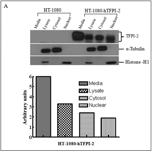
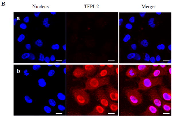
Figure 3. Analyses of TFPI-2 localization in untransfected and stably-transfected HT-1080 cells by immunoblotting and immunocytochemistry.
Wild-type HT-1080 and HT-1080 cells stably-transfected with hTFPI-2 cDNA were cultured under standard growth conditions. (A), For immunoblotting, total lysate and cell fractions were prepared and probed with anti-TFPI-2 antibody as described earlier. The purity of cytosolic and nuclear fractions was verified using anti-alpha-tubulin and anti-histone-H1 antibodies, respectively. For immunoblot analysis, the intensity of blot bands were assessed by densitometric semi-quantitation and depicted by a bar diagram (lower panel). (B), For immunocytochemistry, cells were grown on two-chamber culture slides and incubated with murine monoclonal antibody SK-9 followed by Alexa Fluor-555-conjugated goat anti-mouse IgG as the secondary antibody. The nucleus is counterstained using DAPI in mounting media. (a), HT-1080 cells (b), HT-1080 cells overexpressing TFPI-2.


