Figure 2.

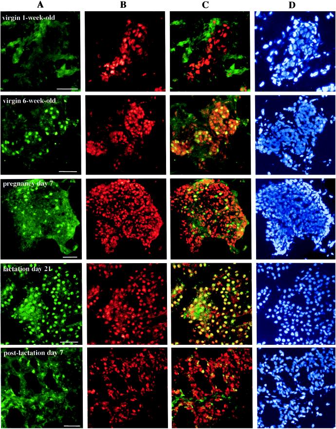
Detection and localization by immunofluorescent labeling of ERα and ERβ proteins in rat mammary glands at different stages of development. Tissue sections from female rats at indicated intervals were stained by sequential exposure of each section to antibodies 6F11, FITC-m, 503, and Cy3-c, followed by DAPI. The green fluorescence spots indicate nuclei containing ERα (A), and the red spots indicate those with ERβ (B). The yellow nuclei observed in overlayered images, especially from lactating glands, indicate that there is coexpression of both receptors in a single epithelial cell (C). Lane D shows the total nuclei in the same section. Pictures are representative of nine slides from three individual rats for each stage. (Bars = 50 μm.)
