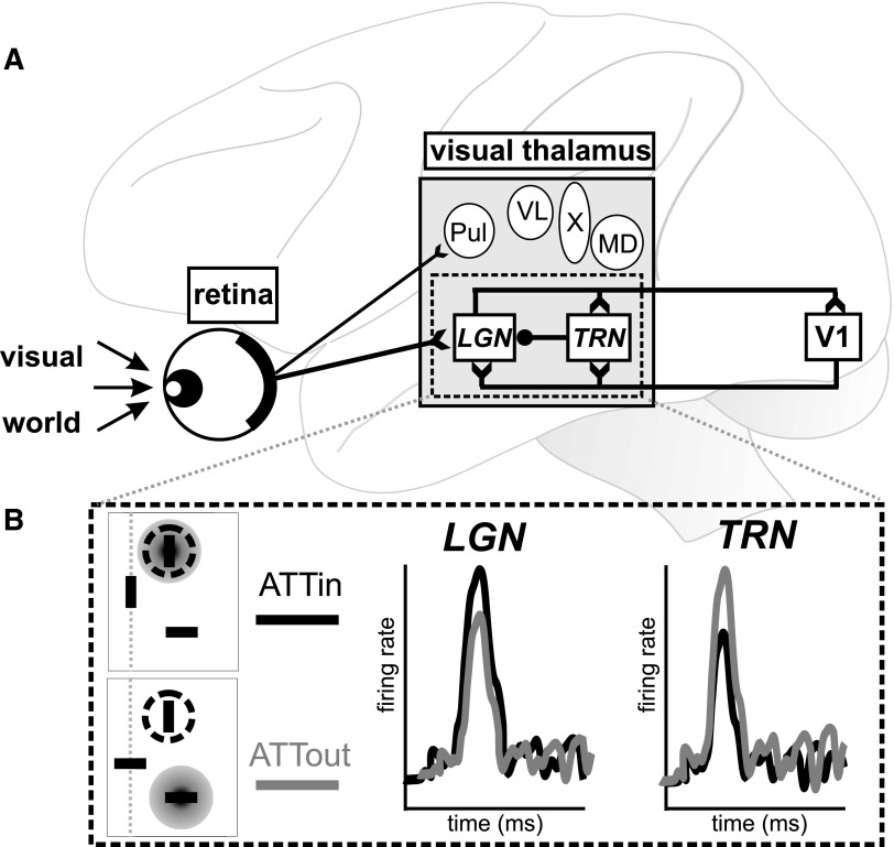
FIG. 1.
Simplified illustration of visual pathway from retina to visual cortex. A: visual information enters the retina and passes through visual thalamus (gray box), including LGN and TRN (italics). Excitatory (open arrow tails) and inhibitory (closed circle) connections are shown for retinal output and the LGN–TRN–V1 circuit. Other nuclei (circles) also likely influence our global visual percept. Note that only Pul and LGN are likely recipients of direct retinal input, as shown, and not all visual nuclei project directly to V1. LGN, lateral geniculate nucleus; MD, mediodorsal nucleus; Pul, pulvinar; TRN, thalamic reticular nucleus; V1, primary visual cortex; VL, ventrolateral nucleus; X, area X. B: key physiological results from McAlonan et al. (2008). Top left: in the ATTin condition, the focus of attention (gray disc) is aligned with the receptive field (dashed circle). Bottom left: in the ATTout condition, attention is fixed on the stimulus outside the receptive field. Right: schematic representation of neuronal responses in LGN and TRN. LGN neurons showed greater activity when attention was focused at the receptive field location (ATTin; black). In contrast, TRN neurons showed relatively larger responses when the focus of attention was outside the receptive field (ATTout; gray).

