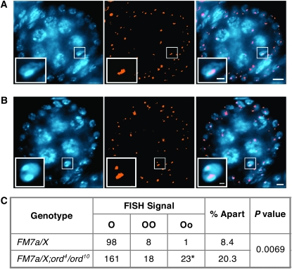
Figure 6.—
Disruption of ord activity weakens FM7a/X heterochromatic pairing. (A and B) Drosophila ovaries from females reared under normal conditions (no aging regimen) were hybridized with the 359-bp repeat pericentric heterochromatin probe (orange) and stained with DAPI (blue). Single egg chambers are shown with the oocyte nucleus enlarged in the inset. Bars, 5 μm in the panels; 1 μm in the insets. (A) Pairing of the distal and the proximal heterochromatin of FM7a with the pericentric heterochromatin of the normal X chromosome results in a single focus within the oocyte nucleus. A stage 3 oocyte is shown. (B) Separated FISH signals result if the interrupted heterochromatin of FM7a is not completely paired with its homolog. A stage 5 oocyte is shown. (C) Heterochromatin pairing was quantified in FM7a/X; ord+ and FM7a/X; ord4/ord10 oocytes using the 359-bp FISH probe. The data represent tabulated results for oocyte stages 2–11 (see supplemental Table S7). “O” denotes a single FISH signal (as seen in A). “OO” denotes separated FISH signals of equal size that occur when the distal heterochromatin of FM7a is not paired with the normal X chromosome. “Oo” denotes separated FISH signals of different sizes (as shown in B) that occur when the centromere-proximal heterochromatin of FM7a fails to pair with the normal X chromosome. (*) 2/23 oocytes contained three FISH signals. Pairing between the heterochromatic regions of the FM7a balancer and a normal X chromosome is disrupted more often in ord4/ord10 than in ord+ oocytes (P = 0.0069).

