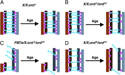
Figure 7.—
Model for how cohesion proteins and pericentric heterochromatin might cooperate to maintain the association of achiasmate homologs. This schematic provides one possible model to explain why heterochromatin-mediated attachments between achiasmate chromosomes depend on sister-chromatid cohesion proteins and how decline of cohesion with age contributes to achiasmate nondisunction. The pericentric heterochromatin of a set of achiasmate homologs is depicted in shades of brown and gray, with sister chromatids shown in different shades. Pink filled circles represent sister-chromatid cohesion proteins. In this model, hypothetical linker proteins that physically connect the two homologs are depicted in blue. Each blue “linker” physically interacts with heterochromatin of one homolog and cohesion proteins that join the two sisters of the other homolog. For simplicity, interaction of the linkers with heterochromatin (vertical blue bars) is shown for only one of the sisters. A more conservative possibility is that the blue “linker” represents a heterochromatin protein on one homolog that interacts with a cohesion protein on the other homolog. (A) In wild-type flies with two normal X chromosomes, even though some deterioration of cohesion occurs during the aging process, the association of achiasmate chromosomes remains intact. (B) In ord4/ord10 oocytes, the centromeric function of ORD is slightly compromised even in young oocytes (fewer filled circles). However, weakening of cohesion with age does not significantly affect the association between homologs when normal amounts of heterochromatin reside near the centromere. (C) In contrast, when centromere-proximal heterochromatin is reduced (FM7a) in ord4/ord10 oocytes, the combination of reduced ORD activity at the centromere, fewer heterochromatin attachment sites and deterioration of cohesion with age causes achiasmate homologs to lose their association. (For ease of illustration, the reduction of heterochromain in FM7a is not drawn to scale). (D) In ord8/ord10 oocytes, the centromeric function of ORD is more severely compromised and age-dependent deterioration of cohesion significantly affects homolog association even when pericentric heterochromatin is normal. Moreover, sister chromatid NDJ is also observed with the stronger ord8 mutation and the frequency of equational NDJ increases when oocytes undergo aging. Note that the loss of homolog association shown in C and D is meant to represent a significant increase in achiasmate missegregation with age, not complete dissociation of the bivalents in 100% of the oocytes.

