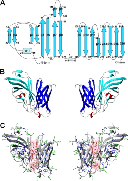
FIGURE 1.
A, topology diagram of the fHbp protein. The α-helices are represented by sky blue cylinders, and the β-strands are cyan arrows. N-term, N terminus; C-term, C terminus. B, ribbon diagram of fHbp. Secondary structure elements are shown. β-strands of the N-terminal domain are shown in cyan and helices are shown in red, whereas β-strands of fHbpC are shown in blue. C, the side chains of the hydrophobic residues involved in interdomains contacts are shown as sticks in red, and pink. Contact surfaces are also reported. The other hydrophobic residues are shown in blue. The charged residues are shown in green. The backbone is shown as a ribbon.

