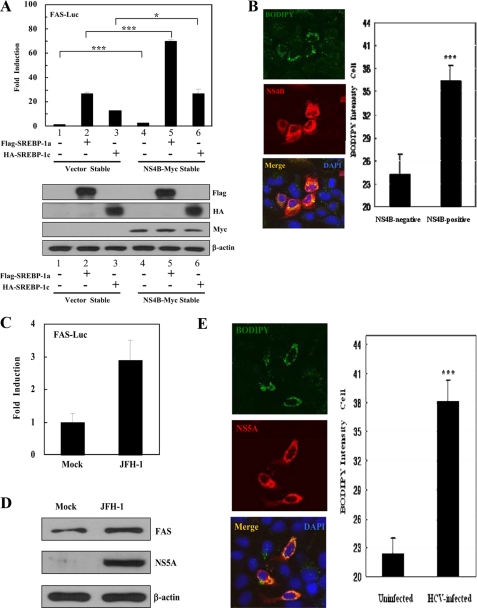
FIGURE 5.
HCV NS4B protein induces lipid accumulation through activation of SREBPs. A, both vector and NS4B-Myc stable cells were cotransfected with FAS-Luc reporter and Flag-SREBP-1a (1–490 amino acids) or HA-SREBP-1c (1–447 amino acids) expression plasmids. At 36 h after transfection, cells were harvested and then luciferase activities were determined (top panel). Data represent the mean of two independent experiments. ***, p < 0.001, vector stable versus NS4B stable cells transfected with Flag-SREBP-1a. *, p < 0.05, vector stable versus NS4B stable cells transfected with HA-SREBP-1c. Equal amounts of cell lysates were subjected to immunoblotting with anti-FLAG, anti-HA, anti-Myc, and anti-β-actin monoclonal antibody (bottom panel). B, Huh7 cells were transfected with NS4B-Myc expression plasmid. At 36 h after transfection, cells were fixed and incubated with anti-Myc monoclonal antibody for 2 h. After being washed with PBS, cells were further incubated with TRITC-conjugated goat anti-mouse IgG and BODIPY 493/503 (1 μm, Invitrogen) for 1 h. Samples were analyzed for immunofluorescence staining using the LSM 510 laser confocal microscopy system and BODIPY intensity was quantified. Each bar represents the average intensity of BODIPY staining. ***, p < 0.001, NS4B-negative cells versus NS4B-positive cells. C, Huh7 cells were either mock-infected or infected with HCV JFH-1. At 3 days after infection, cells were transfected with FAS-Luc reporter plasmid. At 24 h after transfection, cells were harvested and then luciferase activities were determined. D, at 3 days after infection, total cell lysates were immunoblotted with either anti-FAS antibody (top panel) or anti-NS5A antibody (middle panel). Protein expression of β-actin was used as a loading control for the same amount of cell lysates (bottom panel). E, at 3 days after infection, cells were fixed and incubated with anti-NS5A polyclonal antibody for 2 h. After being washed with PBS, cells were further incubated with TRITC-conjugated goat anti-mouse IgG and BODIPY 493/503 (1 μm, Invitrogen) for 1 h. Immunofluorescence staining was performed as described in B. ***, p < 0.001, mock-infected cells versus HCV-infected cells. Cells were counterstained with 4′,6-diamidino-2-phenylindole (DAPI) to label nuclei.

