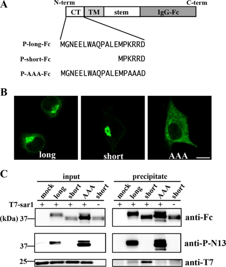
FIGURE 5.
Interaction between chimeric GlcAT-Ps and Sar1. A, schematic diagrams of chimeric GlcAT-Ps are shown. The catalytic domains of these chimeras were replaced with the human IgG-Fc region. The N-terminal cytoplasmic tails of three chimeras, P-long-Fc, P-short-Fc, and P-AAA-Fc, correspond to those of full-length lGlcAT-P, sGlcAT-P, and lGlcAT-P-AAA, respectively. B, Neuro2A cells expressing P-long-Fc (left), P-short-Fc (middle), or P-AAA-Fc (right) were immunostained with anti-human IgG-Fc pAb. Note that the staining patterns shown here resemble those of the full-length enzymes (Figs. 2A and 3B). Bar, 10 μm. C, Neuro2A cells were transfected with the P-long-Fc, P-short-Fc, or P-AAA-Fc expression plasmid or the empty plasmid (mock). Cells were lysed and incubated with recombinant T7-tagged Sar1, followed by incubation with Protein G beads. The lysate mixture before precipitation (left) and the proteins precipitated with the beads (right) were Western blotted with anti-human IgG-Fc pAb (top), anti-P-N13 pAb (middle), or anti-T7 mAb (bottom).

