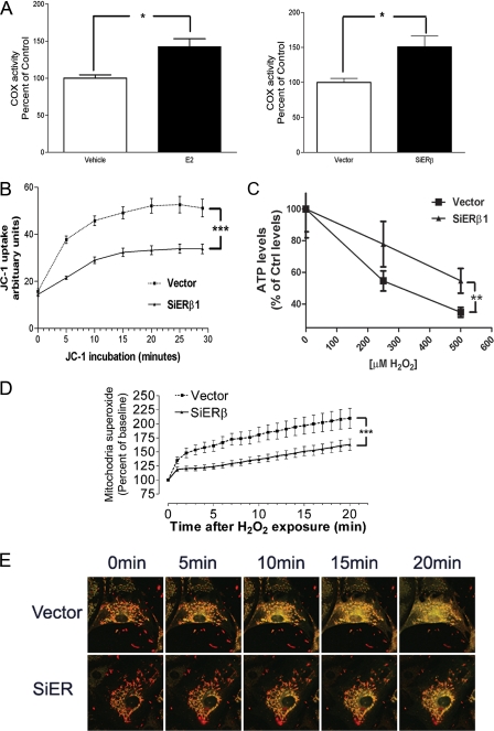
FIGURE 3.
A, right panel, 17β-estradiol (E2) treatment increases cytochrome c oxidase activity in HT-22 cells. *, p < 0.05 versus vehicle. Left panel, quantitative analysis of cytochrome c oxidase activity in vector and siERβ cells. *, p < 0.05 versus vector. B, quantitative analysis of dynamic change of JC-1 uptake in vector-transfected (Vector) and ERβ knockdown HT-22 cells (siERβ). n = 16. ***, p < 0.001 versus vector. C, H2O2-induced decrease of ATP production in vector-transfected and ERβ knockdown HT-22 cells. n = 6. **, p < 0.01 versus vector. D, quantitative analysis of dynamic changes in H2O2-induced mitochondrial superoxide production, measured by MitoSox fluorescence, in vector and siERβ HT-22 cells. n = 16. ***, p < 0.001 versus vector. E, effect of ERβ on H2O2-induced Δψm collapse in HT-22 cells. Confocal microscopy images show the same field of cells viewed before (0 min) and at 5, 10, 15, and 20 min after H2O2 (1 mm) exposure in vector and siERβ HT-22 cells.

