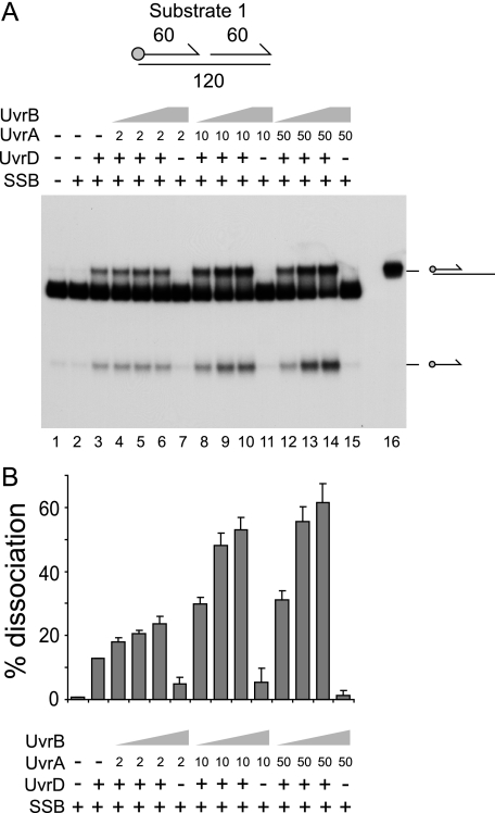
FIGURE 1.
UvrA and -B stimulate UvrD-catalyzed unwinding of a nicked duplex. A, unwinding of substrate 1 (numbers indicate the length in base pairs of each duplex) in the presence of SSB (125 nm), UvrD (10 nm), UvrA (nm concentrations shown), and UvrB (10 nm in lanes 4, 8, and 12, 100 nm in lanes 5, 9, and 13, and 500 nm in lanes 6, 7, 10, 11, 14, and 15). Lane 16 contained a partial duplex marker. In the substrate diagram, the circle represents the position of the 5′ 32P label, whereas arrows represent the 3′ ends of oligonucleotides. B, degree of unwinding of substrate 1 in lanes 2-15. Data represent the means of two experiments.

