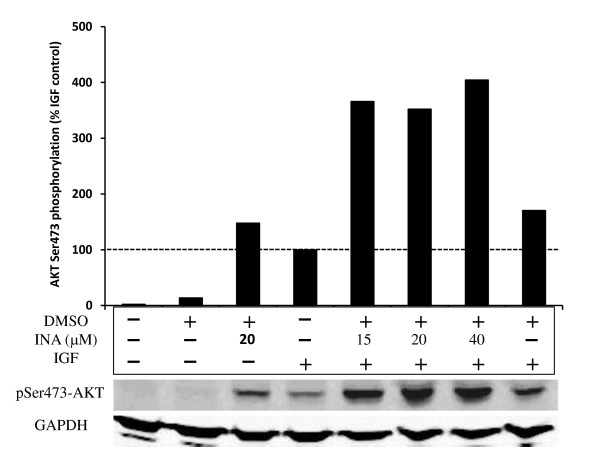
Figure 8.
INA-UV treatment does not inhibit IGF1 signaling. Akt phosphorylation was assessed by Western detection and quantified. The different samples correspond to MCF7 cells untreated, exposed or not to 1% DMSO, INA at the indicated concentrations and/or IGF1. All the samples exposed to DMSO or INA were irradiated with UV. The Akt phosphorylation signal was normalized to the signal obtained with MCF7 exposed only to IGF1. The Western signal obtained with GAPDH on the same samples is shown for loading control.

