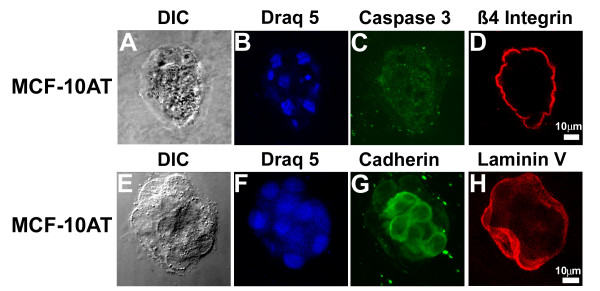
Figure 9.
Altered morphology of MCF-10AT cells in grown for 20 days in embedded three-dimensional culture. Differential interference contrast (DIC) images (A, E) show that MCF-10AT cells grown under these conditions form irregular acini but not the multi-acinar structures seen in overlay cultures (see Figure 2). Nuclei were stained with Draq5 (B, F). Apoptosis, cell:cell junctions, and basement membrane formation were assessed by confocal microscopy with caspase-3 (C), cadherin (G), β4 integrin (D), and Laminin V (H) staining. Scale bars, 10 μm.

