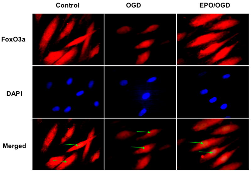
Figure 1. Erythropoietin (EPO) excludes FOXO3a from nuclear translocation during oxidative stress.
Application of EPO (10 ng/ml) during an 8 hour period of oxygen-glucose deprivation (OGD), OGD alone, or in untreated primary rat cerebral endothelial cells (Control) was followed at 6 hours with immunofluorescent staining with primary rabbit anti-FoxO3a antibody then by Texas red conjugated anti-rabbit secondary antibody. Nuclei of endothelial cells were counterstained with DAPI and are highlighted in the bottom panels for Control, OGD, and EPO/OGD with green arrows. In merged images, endothelial cells with combined EPO and OGD demonstrate minimal FoxO3a staining in the nuclei of cells (white) and show the cytoplasm of endothelial cells with significant FoxO3a staining (red). These observations are in contrast to cells with OGD alone with significant FoxO3a staining in both the cytoplasm and the nuclei of endothelial cells, demonstrating the ability of EPO to prevent the translocation of FoxO3a to the nucleus to initiate a pro-apoptotic program.

