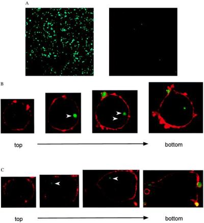
Figure 1.

Platelet binding to LOX-1-expressing cells. (A) Association of platelets labeled with calcein (green) was observed in BLOX-1-CHO (Left) but not in CHO-K1 (Right). (B and C) The fate of platelets associated with BLOX-1-CHO (B) or BAE (C) was observed under a confocal laser microscope. The cell surface was visualized with rhodamine-labeled Con A (red), and platelets were labeled with calcein (green). The surfaces of the platelets outside of BLOX-1-CHO and BAE show signals of rhodamine. Platelets inside of the cells do not (arrowheads). The platelet signals are detected in the intermediate layers of slices but not in upper or lower slices.
