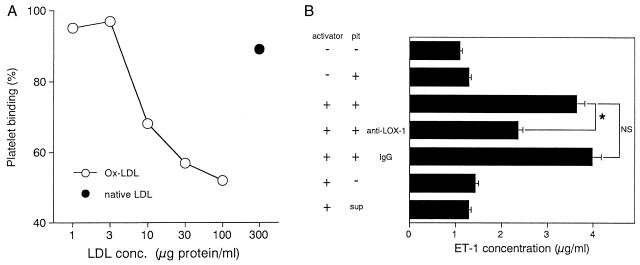
Figure 4.
(A) OxLDL dose-dependently inhibited the binding of platelets to BLOX-1-CHO cells, but native LDL did not. The cells were viable even at the highest concentration of OxLDL, being observed with light microscopy. The values are expressed as a percentage of the platelet binding without supplementation of lipoproteins. (B) BAE were incubated for 3 h with the resting or activated platelets, which were stimulated by thrombin (1 unit/ml) and collagen (20 μg/ml). The amount of ET-1 released from BAE in the next 20 h was determined by enzyme immunoassay. The anti-LOX-1 antibody blocked a significant part of the ET-1 release enhanced by activated platelets. Note that the supernatant of activated platelets (sup) had limited effects in comparison with whole platelets. Data represent the means ± SEM of triplicate experiments. Asterisks indicate a significant difference (P < 0.05). plt, platelet; NS, not significant.

