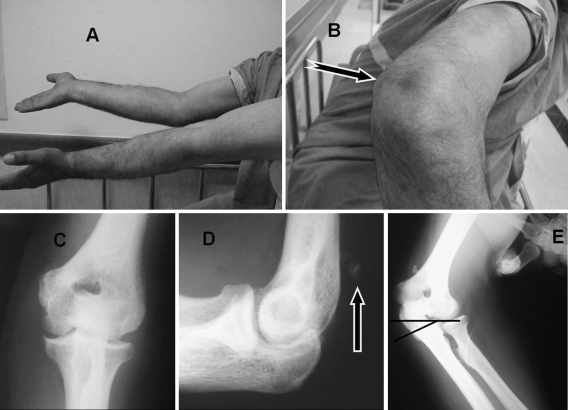
Fig. 1.
Preoperative clinical appearance of the patient (a, b). Note the extension loss at the involved right elbow (a) and dimple (arrow) at the posterior aspect of the elbow (b). Direct radiographs anteroposterior (c) and lateral (d). Lateral radiograph shows ‘fleck sign’ (arrow) on posterior aspect of the distal humerus (d). Significant medial opening of the ulno-humeral joint (e) with application of valgus stress (black lines represent humeral and ulnar joint lines)

