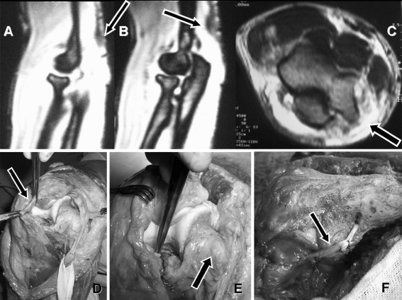
Fig. 2.
Preoperative oblique sagital magnetic resonance images showing ruptured triceps tendon (arrow on a) and avulsed bony fragment (arrow on b). Transverse MRI shows increased intensity surrounding the medial epicondyle (c) indicating injury to neighboring structures. Clinical photographs that are taken during the surgery show thick fibrous healing tissue (arrow on d). Medial epicondyle is devoid of any tissue (arrow on e) including UCL and FPMG. Reconstruction of UCL (arrow on f) is performed free palmaris longus tendon autograft

