
Fig. 2.
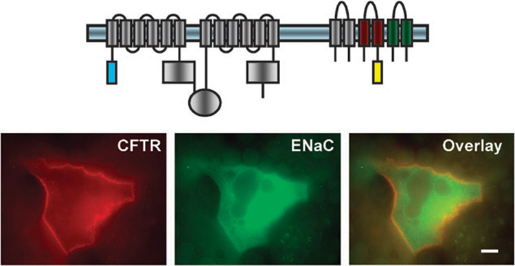
Co-localization of ECFP-CFTR and β-YFP-αγ-ENaC at the plasma membrane of the MDCK cells. Fluorescently tagged CFTR and ENaC constructs were transfected into MDCK cells using Lipofectamine2000. Images were captured with an Olympus IX70 inverted epifluorescence microscope equipped with a 100× oil objective with a numerical aperture of 1.4. Scale bar 10 µm. Reproduced with permission from Berdiev et al., 2007, The American Society for Biochemistry and Molecular Biology.74