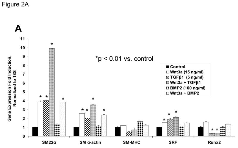
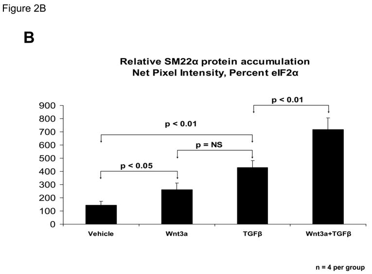
Figure 2. Wnt3a and TGFβ1 signals augment SM22α expression in C3H10T1/2 cells.
C3H10T1/2 cells were treated for 4 days with either vehicle, 15 ng/ml Wnt3a, 5 ng/ml TGFβ1, and/or 100 ng/ml BMP2 in the combinations indicated. Total cellular RNA was extracted and gene expression analyzed by RT-qPCR as described in “Materials and Methods.” Wnt5a (15 ng/ml) was unable to replace Wnt3a actions in this assay (data not shown). Panel B, quantitative western blot analysis confirms that Wnt3a is capable of significantly augmenting SM22α protein accumulation in either the presence or absence of TGFβ1. Data were obtained from quantitative image analysis of 4 independent replicates following 4 days of the indicated treatments, normalizing the SM22α signal to eIF2alpha signal in each sample.


