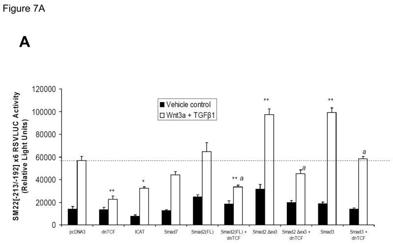
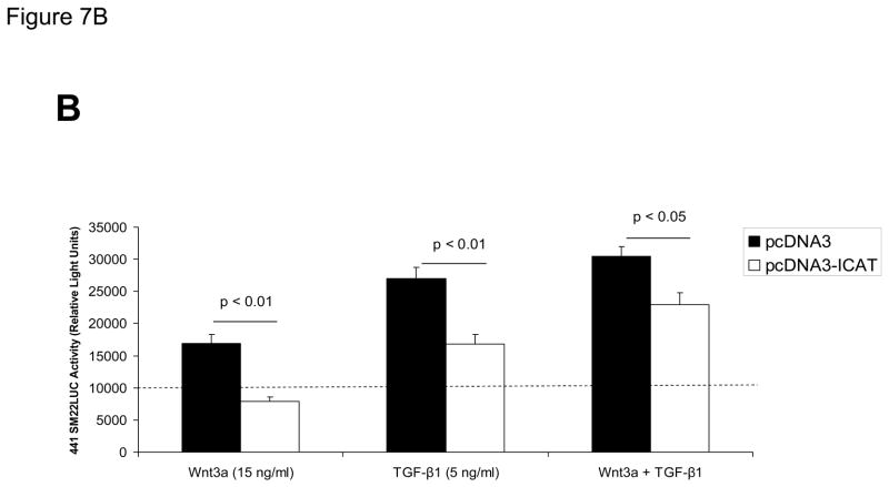
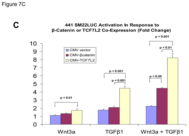
Figure 7. Intracellular antagonists and agonists of canonical Wnt/β-catenin signaling regulate transcriptional activation of SM22[−213/−192]-RSVLUC.
Panel A, expression plasmids for dominant negative TCF (dnTCF), ICAT, and Smad7 were co-transfected with SM22[−213/−192]×6-RSVLUC, and the impact on Wnt3a+TGFβ1 induction examined as done in Figure 3. Note Smad2Δexon3 and Smad3 significantly augmented Wnt3a+TGFβ1 stimulation, and that dnTCF expression consistently suppressed activation. See text for details. **, p < 0.001 vs. Wnt3a+TGFβ1 stimulated pcDNA3 control (dotted line), by post-hoc Tukey’s test following an ANOVA with p < 0.0001. *, p < 0.01 vs. Wnt3a+TGFβ1 stimulated pcDNA3 control. a, p < 0.001 vs. the corresponding Smad co-transfection that lacks dnTCF. Panel B, ICAT, the inhibitor of β-catenin and TCF, reduced Wnt3a and TGFβ1 induction of SM22α activity in the native promoter context of 441 SM22LUC. Panel C, 441 SM22LUC was upregulated by transient co-expression of either β-catenin or TCF7L2, the former observed in the presence of concomitant Wnt3a+TGFβ1 treatment.


