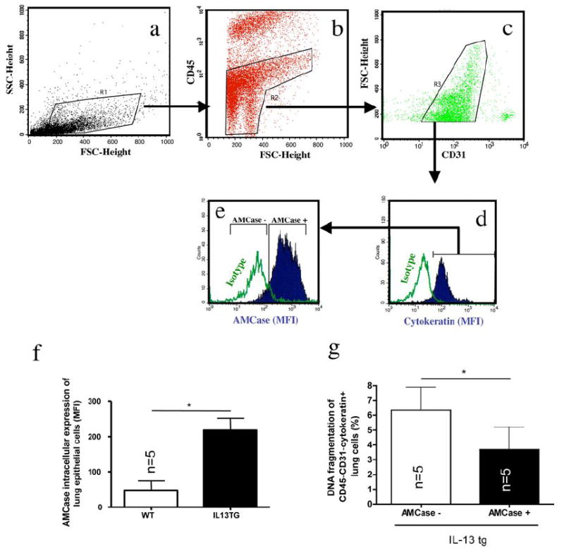
Figure 2. In vivo analysis of AMCase in lung epithelial cells.

a-e: Lung epithelial cells were characterized according to the following gating algorithm: Within digested/strained and red cell and macrophage-depleted whole lung cell suspensions (see for details Materials and Methods section), non-debris cells were gated (a) and hematopoietic (CD45+) cells were further excluded (b). Within CD45- negative lung cells CD31+ endothelial cells were excluded (c). Within the CD45-CD31- lung cell population, cells positive for intracellular pan-Cytokeratin (d) were considered as airway epithelial cells according to a modified method as described previously [64;65]. Intracellular AMCase expression was robustly detectable in pan-Cytokeratin+ cells (e).
f. Intracellular AMCase was stained in permeabilized pan-Cytokeratin+ epithelial cells in wildtype (WT) and IL-13 transgenic overexpressing (IL-13 TG). * p<0.05, Mann-Whitney U test
g. Airway epithelial cell apoptosis was assessed using DNA fragmentation (FACS TUNEL staining) in IL-13 tg mice in CD45-CD31-Cytokeratin+ lung cells with high (AMCase MFI>200, AMCasehigh) or low (AMCase MFI <200, AMCaselow) intracellular AMCase expression. * p<0.05, Mann-Whitney U test
